


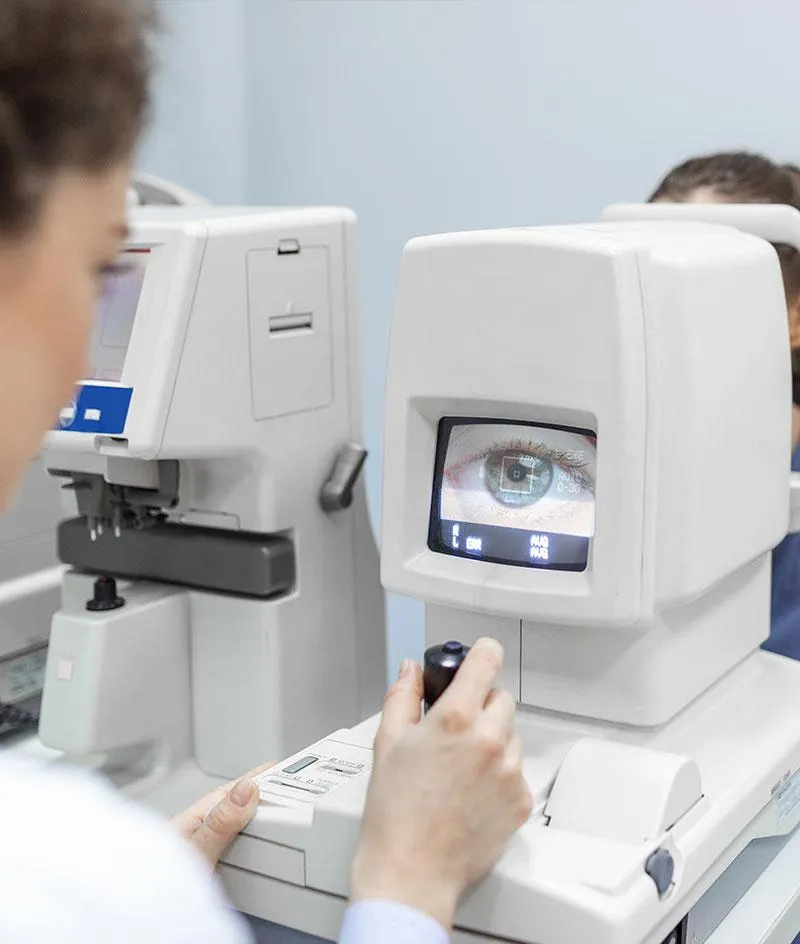
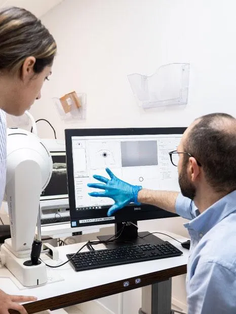
Eye Diagnostics and Imaging
The Harley Street Eye Centre uses a comprehensive imaging system that has been seamlessly integrated with the advanced technology and capabilities of the Carl Zeiss Forum platform.
This integration brings together state-of-the-art imaging equipment and software solutions developed by Carl Zeiss, a renowned leader in the field of medical optics and imaging.
SINCE 2019, OUR EYE CLINIC HAS TREATED OVER 4,700 PATIENTS WITH ADVANCED EYE CARE.
What Can The Harley Street Eye Centre Do For Your Corneal Scratch?
Corneal Abrasion Treatment: A clinical corneal consultation will be needed, where we will apply an eye drop to stain the eye and confirm the scratch. Scratches in the cornea tend to be in the epithelial layer (top layer of the cornea) and thus future regrowth is expected however this will take a little time.
We can assist regrowth by placing a clear bandage contact lens on to protect the cornea for a few days while regrowth occurs.
You will also be given a topical eye antibiotic to prevent infection from bacteria that may have been introduced.

Does any of this sound like YOU?
You need frequent changes in your prescription eyeglasses or contact lenses?
You're finding it increasingly difficult to achieve clear vision with corrective lenses?
You've got poor night vision or increased sensitivity to glare?
You've got distorted or blurred vision, especially in one eye?
You often see halos or ghosting around lights?
You experience eye strain or eye fatigue, particularly after visual tasks?
Your contact lenses are becoming uncomfortable or even difficult to wear?
You've already tried a lot of non-surgical treatments to help improve your vision but they have all failed?

Mr Marwan Ghabra
Clinical Director
It is important to note that these signs are not definitive indicators of keratoconus or the need for surgery. After a thorough eye examination and the appropriate diagnostic tests, a qualified eye care professional at The Harley Street Eye Centre can determine a proper diagnosis and treatment plan, if required.
Imaging Suite fully integrated by
Carl Zeiss Forum
The Imaging Suite encompasses a range of cutting-edge imaging modalities and devices designed to provide precise and detailed visualization of ocular structures and conditions. These may include:
These may include:
Optical Coherence Tomography (OCT): OCT is a non-invasive imaging technique that utilizes light waves to capture high-resolution cross-sectional images of the retina and other ocular structures. It aids in the diagnosis and monitoring of various eye conditions, such as macular degeneration, diabetic retinopathy, and glaucoma.
Fundus Photography: Fundus photography involves capturing high-resolution images of the back of the eye, specifically the retina, blood vessels, and optic nerve. It provides a valuable tool for documenting and monitoring changes in the eye over time, aiding in the diagnosis and management of retinal diseases.
Anterior Segment Imaging: This includes technologies such as anterior segment OCT, which allows for detailed imaging of the front portion of the eye, including the cornea, iris, and anterior chamber. It assists in the assessment of corneal conditions, anterior segment abnormalities, and the planning of corneal surgeries.
Corneal Topography: Corneal topography is a diagnostic tool used to map the shape and curvature of the cornea. It provides essential information for the evaluation of corneal conditions, such as keratoconus, corneal irregularities, and preoperative planning for refractive surgeries.
Biometry: Biometry is utilized in cataract surgery planning to accurately measure the size and shape of the eye, specifically the length of the eyeball and the curvature of the cornea. This information helps determine the appropriate power of the intraocular lens (IOL) to be implanted during cataract surgery.
By integrating these imaging modalities into the Carl Zeiss Forum platform, healthcare professionals gain a powerful and comprehensive suite of tools for diagnosing, monitoring, and planning treatment for various ocular conditions.
The seamless integration ensures smooth data transfer, enhanced analysis capabilities, and efficient workflow management, enabling eye care providers to make well-informed decisions and deliver optimal patient care.
Got a Question?
Ask Mr Ghabra & His Team Of Doctors
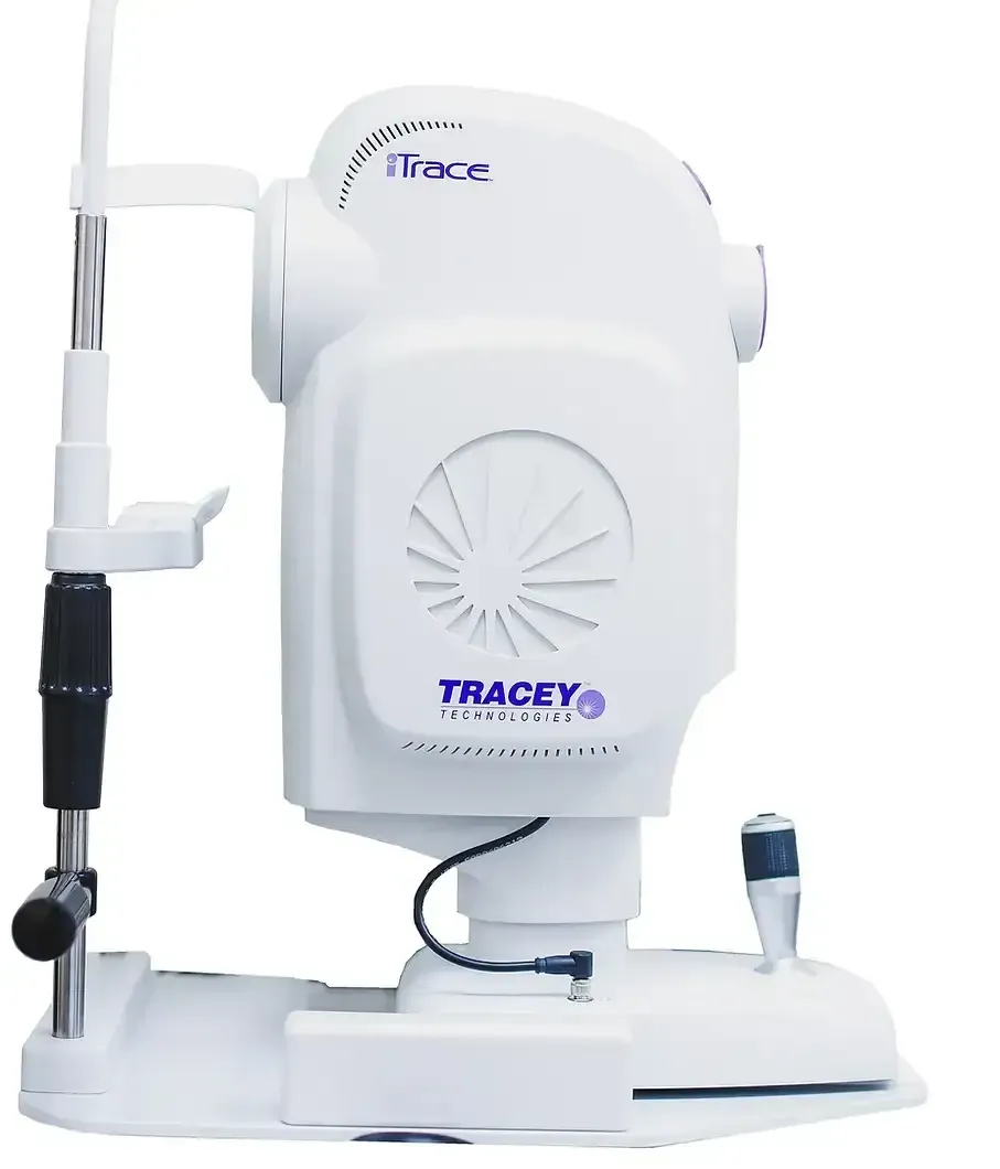
I-Trace Aberrtometry
I-TRACE Aberrometry is an advanced diagnostic tool used in ophthalmology to measure and analyze the optical aberrations of the eye.
It provides a comprehensive assessment of the eye's refractive errors and irregularities, including higher-order aberrations that may not be detected by traditional methods.
Benefits of The I-TRACE Aberrometry
Precise Measurement: I-TRACE Aberrometry offers highly precise and accurate measurements of the eye's optical aberrations. It provides detailed information about irregularities in the cornea, lens, and other structures of the eye, enabling a more comprehensive understanding of the patient's visual system.
Personalized Treatment: By obtaining detailed aberration data, I-TRACE Aberrometry allows eye care professionals to create personalized treatment plans. The information helps in determining the most suitable corrective measures, such as customized contact lenses, wavefront-guided LASIK, or other vision correction procedures tailored to the specific needs of the patient.
Detection of Irregularities: I-TRACE Aberrometry can detect and quantify subtle irregularities in the eye that may contribute to visual disturbances. It identifies higher-order aberrations that are beyond the scope of conventional refractive assessments, offering a more comprehensive evaluation of the patient's visual system.
Refractive Surgery Planning: For patients considering refractive surgery, I-TRACE Aberrometry plays a crucial role in assessing their candidacy and determining the optimal treatment approach. The detailed aberration measurements help in planning and performing procedures like wavefront-guided LASIK or PRK, resulting in improved outcomes.
Monitoring Progress: I-TRACE Aberrometry can be used to monitor the progress of vision correction treatments or surgeries over time. By periodically assessing aberration patterns, eye care professionals can track changes in the visual system and make necessary adjustments or enhancements if required.
Research and Advancements: I-TRACE Aberrometry contributes to ongoing research and advancements in the field of ophthalmology. The data collected from patients using this technology aids in understanding visual abnormalities, developing new treatment techniques, and refining existing procedures to enhance patient outcomes.
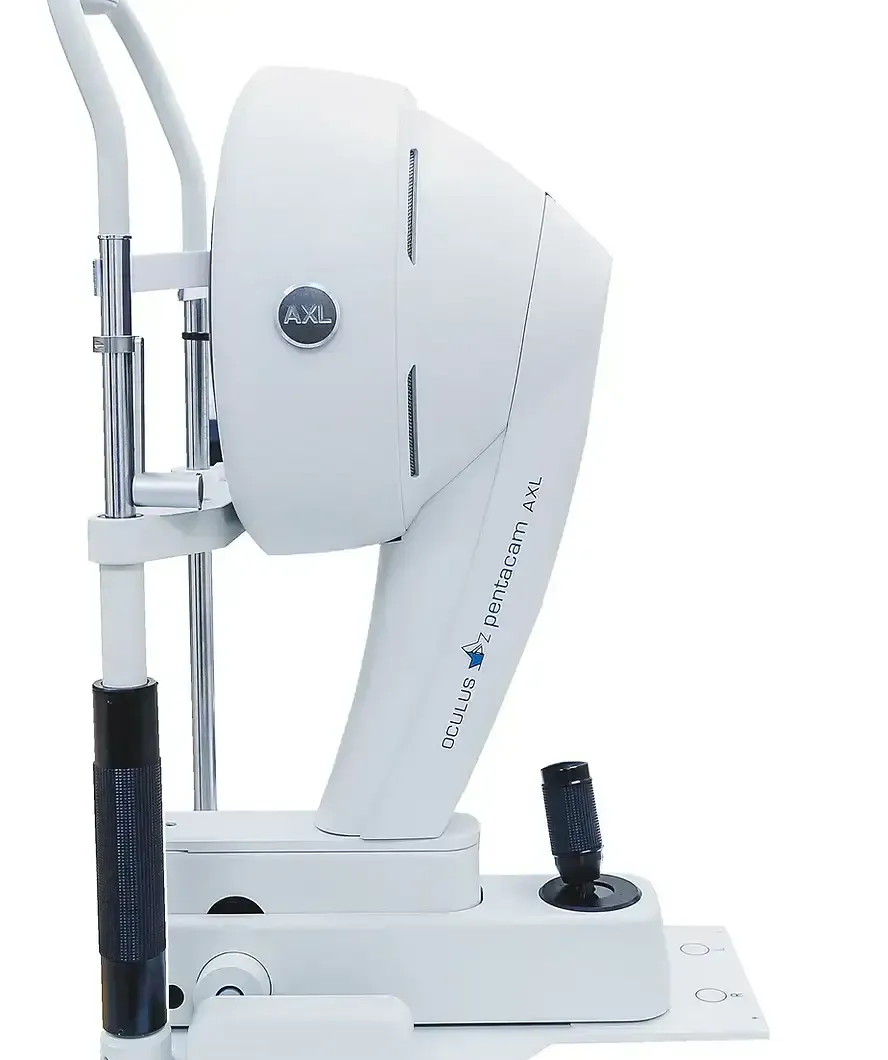
Oculus AXL Pentacam
The Oculus AXL Pentacam is a state-of-the-art imaging device used in ophthalmology for comprehensive corneal analysis.
It combines Scheimpflug imaging and optical biometry to provide detailed measurements and imaging of the anterior segment of the eye, including the cornea, anterior chamber, and crystalline lens.
Benefits of The Oculus AXL Pentacam
Precise Corneal Assessment: The Oculus AXL Pentacam offers precise and detailed measurements of the cornea, including its curvature, thickness, and shape. This information is crucial for diagnosing and monitoring various corneal conditions such as keratoconus, corneal ectasia, and corneal dystrophies.
Detection of Irregularities: The device can detect subtle corneal irregularities that may not be apparent in conventional examinations. It provides valuable insights into corneal aberrations, asymmetries, and other irregularities, helping in the early detection and management of corneal disorders.
Pre-operative Planning for Refractive Surgery: The Oculus AXL Pentacam plays a vital role in pre-operative planning for refractive surgeries like LASIK, PRK, or implantable collamer lenses. It provides comprehensive corneal measurements, allowing surgeons to assess corneal thickness, shape, and curvature to determine the suitability of patients for different refractive procedures.
Customised Treatment Approaches: With the detailed corneal data provided by the Oculus AXL Pentacam, eye care professionals can tailor treatment approaches to individual patients. It helps in selecting the appropriate type of refractive surgery, determining the ideal ablation patterns, and optimizing treatment outcomes for each patient's unique corneal characteristics.
Evaluation of Lens Parameters: In addition to corneal assessment, the Oculus AXL Pentacam also provides accurate measurements of the crystalline lens. This information is valuable for determining the power and position of intraocular lenses (IOLs) in cataract surgery and other lens-related procedures.
Progress Monitoring: The device allows for the tracking of corneal changes over time. By comparing sequential measurements, eye care professionals can assess the stability of corneal conditions, evaluate the effectiveness of treatment interventions, and make informed decisions regarding ongoing management.
Research and Advancements: The data collected from the Oculus AXL Pentacam contributes to ongoing research and advancements in corneal and refractive surgery fields. The device aids in the development of new diagnostic techniques, treatment strategies, and understanding of corneal biomechanics.

Carl Zeiss Angio OCT
The Carl Zeiss Angio OCT (Optical Coherence Tomography) is an advanced imaging technology used in ophthalmology to visualize and evaluate the blood vessels and structures of the eye.
It combines the capabilities of OCT imaging with angiography, allowing for non-invasive, high-resolution imaging of ocular vasculature.
Benefits of The Carl Zeiss Angio OCT
Visualization of Retinal Vasculature: The device provides detailed imaging of the retinal blood vessels, allowing for the visualization and assessment of the retinal circulation. This helps in the diagnosis and management of various retinal vascular conditions such as diabetic retinopathy, macular degeneration, and retinal vein occlusions.
Detection of Pathological Changes: The Carl Zeiss Angio OCT can detect subtle changes in the retinal vasculature, including microaneurysms, neovascularization, and areas of non-perfusion. These findings are crucial for the early detection and monitoring of retinal diseases, enabling timely intervention and treatment.
Assessment of Choroidal Vasculature: In addition to the retina, the device allows for the evaluation of the choroidal vasculature, which plays a vital role in maintaining retinal health. The imaging capabilities of the Carl Zeiss Angio OCT help in identifying choroidal abnormalities and understanding their impact on retinal function.
Quantitative Analysis: The Carl Zeiss Angio OCT provides quantitative measurements of various parameters related to retinal and choroidal vasculature, such as vessel density, flow rates, and perfusion indices. These measurements aid in objective evaluation, disease staging, and monitoring treatment response.
Non-Invasive Imaging: The Carl Zeiss Angio OCT is a non-invasive imaging technique that allows for high-resolution visualization of ocular structures without the need for contrast agents or invasive procedures. This enhances patient comfort and safety during the examination.
Guiding Treatment Decisions: The detailed imaging and information obtained from the Carl Zeiss Angio OCT assist in guiding treatment decisions. It helps ophthalmologists determine the most appropriate therapeutic interventions, such as anti-vascular endothelial growth factor (anti-VEGF) injections, laser treatments, or surgical procedures, based on the specific vascular characteristics observed.
Research and Advancements: The data collected through the Carl Zeiss Angio OCT contributes to ongoing research and advancements in the field of ophthalmology. It aids in the development of new diagnostic criteria, treatment algorithms, and understanding of ocular vascular diseases.
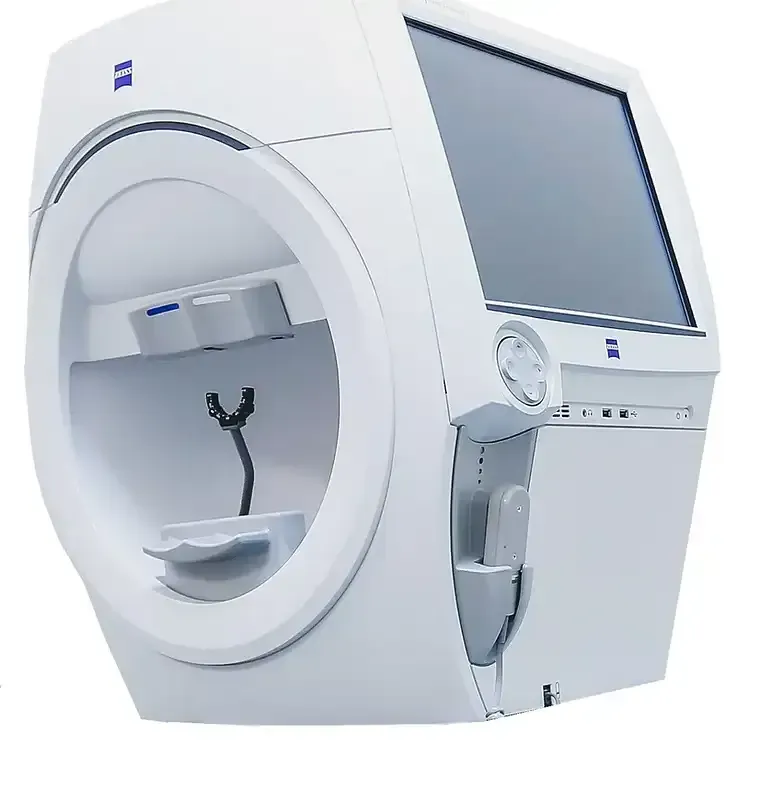
Carl Zeiss Humphrey
Carl Zeiss Humphrey is a brand of advanced diagnostic equipment used in ophthalmology. It is primarily known for its Humphrey Visual Field Analyzer, a device that assesses a patient's visual field.
This tool is essential in diagnosing and managing various eye conditions, such as glaucoma, optic nerve damage, and neurological disorders affecting vision.
Benefits of The Carl Zeiss Humphrey
Accurate diagnosis: The Humphrey Visual Field Analyzer provides precise and detailed measurements of a patient's visual field, enabling eye care professionals to make accurate diagnoses of conditions such as glaucoma, optic nerve damage, and neurological disorders affecting vision.
Disease management: By regularly monitoring changes in a patient's visual field over time, the Humphrey Visual Field Analyzer aids in effectively managing and treating eye diseases. It helps eye care practitioners determine the progression of conditions and assess the efficacy of treatment plans.
Early detection: The device is highly sensitive in detecting early signs of visual field abnormalities, allowing for early intervention and timely treatment. This is particularly important in conditions like glaucoma, where early detection and treatment can significantly slow down disease progression.
Customizable testing options: The Humphrey Visual Field Analyzer offers a range of testing protocols and parameters that can be customized to suit the specific needs of individual patients. This flexibility allows for comprehensive assessments tailored to the patient's condition and provides reliable data for informed decision-making.
Patient-friendly experience: Carl Zeiss Humphrey equipment is designed to ensure patient comfort and ease of use. The testing process is non-invasive, and the device incorporates intuitive user interfaces and ergonomic features, enhancing the overall patient experience during visual field examinations.
Widely accepted and trusted: Carl Zeiss Humphrey equipment is widely recognized and respected in the field of ophthalmology. It has been extensively validated through scientific research and is trusted by eye care professionals worldwide for its accuracy, reliability, and effectiveness in diagnosing and managing various eye conditions.
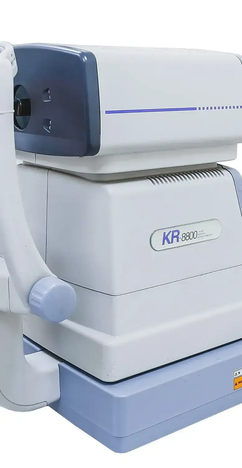
Auto-Keratometry
Auto-Keratometry is a diagnostic technique used in optometry and ophthalmology to measure the curvature of the cornea, which is the clear, front part of the eye.
Benefits of The Auto-Keratometry
Corneal curvature assessment: Auto-Keratometry provides accurate and objective measurements of the corneal curvature, aiding in the diagnosis and monitoring of various corneal conditions, such as astigmatism, keratoconus, and corneal irregularities.
Contact lens fitting: Auto-Keratometry plays a crucial role in fitting contact lenses. By determining the precise corneal curvature, eye care practitioners can select the appropriate contact lens parameters, ensuring a proper fit and optimal vision correction.
Refractive surgery planning: Prior to refractive surgeries like LASIK or PRK, Auto-Keratometry helps determine the corneal shape and curvature, guiding the surgeon in determining the treatment plan, calculating the amount of tissue to be removed, and ensuring the desired outcome.
Monitoring corneal changes: Auto-Keratometry enables practitioners to monitor changes in corneal curvature over time. This is particularly important in conditions like keratoconus, where progressive corneal steepening can be assessed, and treatment interventions can be initiated at appropriate stages.
Diagnostic tool: Auto-Keratometry is an essential diagnostic tool for assessing the health of the cornea. It helps in identifying corneal irregularities, corneal dystrophies, and other abnormalities that may affect visual acuity and require further investigation or treatment.
Quick and non-invasive procedure: Auto-Keratometry is a rapid and non-invasive procedure that provides immediate results. It is a well-tolerated test for patients, requiring minimal patient cooperation and providing valuable information in a relatively short amount of time.
E-Swin IPL
E-Swin IPL (Intense Pulsed Light) is a technology used in medical aesthetics and dermatology for various skin treatments.
Ideal for the treatment of:
Dry Eye
Meibomian Gland Dysfunction (MGD)
Rosacea

Benefits of The E-SWIN IPL
Broad range of treatments: E-Swin IPL offers a versatile platform for addressing a wide range of skin concerns. It can effectively treat conditions such as unwanted hair, pigmentation issues, vascular lesions, acne, rosacea, and signs of aging like fine lines and wrinkles.
Non-invasive procedure: E-Swin IPL treatments are non-invasive, meaning they do not require surgical incisions. This makes it a safe and comfortable option for patients seeking skin rejuvenation or hair removal without the risks and downtime associated with invasive procedures.
Precise and targeted treatment: The technology of E-Swin IPL allows for precise targeting of specific areas or concerns on the skin. The device emits a broad spectrum of light that can be adjusted to the desired wavelength, enabling practitioners to deliver focused treatments and achieve optimal results.
Customizable settings: E-Swin IPL devices are equipped with customizable settings, allowing practitioners to tailor treatments to the unique needs of each patient. This flexibility ensures personalized treatment plans, taking into account skin type, condition severity, and patient comfort.
Effective and long-lasting results: E-Swin IPL treatments have been shown to deliver significant and long-lasting results. For example, in hair removal, the light energy targets hair follicles, leading to a reduction in hair growth over time. Similarly, treatments for pigmentation issues or vascular lesions can result in improved skin tone and texture with lasting effects.
Safe and well-established technology: E-Swin IPL devices are widely used in the medical aesthetics industry and have a proven safety record when used by trained professionals. They undergo rigorous testing and meet strict regulatory standards to ensure patient safety and treatment efficacy.

Just 8 Reasons To Choose The Harley Street Eye Centre
Expertise and Experience: - The Harley Street Eye Centre boasts a team of highly skilled and experienced eye specialists who are renowned in their respective fields. They have the knowledge and expertise to provide top-quality eye care and treatment.
State-of-the-Art Facilities: - The clinic is equipped with advanced and cutting-edge technology, ensuring that patients receive the most accurate diagnoses and effective treatments available in the field of ophthalmology.
Comprehensive Eye Care - The Harley Street Eye Centre offers a wide range of eye care services, including routine eye exams, refractive surgery (such as LASIK), cataract surgery, corneal transplants, glaucoma treatment, and more. Patients can receive comprehensive eye care under one roof.
Personalised Treatment Plans - Each patient is unique, and at The Harley Street Eye Centre, the treatment plans are tailored to meet individual needs. The doctors take the time to understand the specific concerns of each patient and develop personalised treatment strategies accordingly.
Strong Reputation - The Harley Street Eye Centre has built a strong reputation over the years due to its commitment to excellence. The clinic has a track record of successful treatments and satisfied patients, which is reflected in positive reviews and testimonials.
Commitment to Excellence - The Harley Street Eye Centre has built a strong reputation over the years due to its commitment to excellence. The clinic has a track record of successful treatments and satisfied patients, which is reflected in positive reviews and testimonials.
Collaborative Approach - The Harley Street Eye Centre is dedicated to providing the highest standard of eye care and treatment. They prioritise patient safety, satisfaction, and long-term visual health, making them a trusted choice for individuals seeking the best eye clinic for their needs.
Continuing Education and Research - The team at The Harley Street Eye Centre remains at the forefront of advancements in ophthalmology through continuous education and involvement in research. This commitment to staying updated with the latest developments ensures that patients receive the best available treatments.
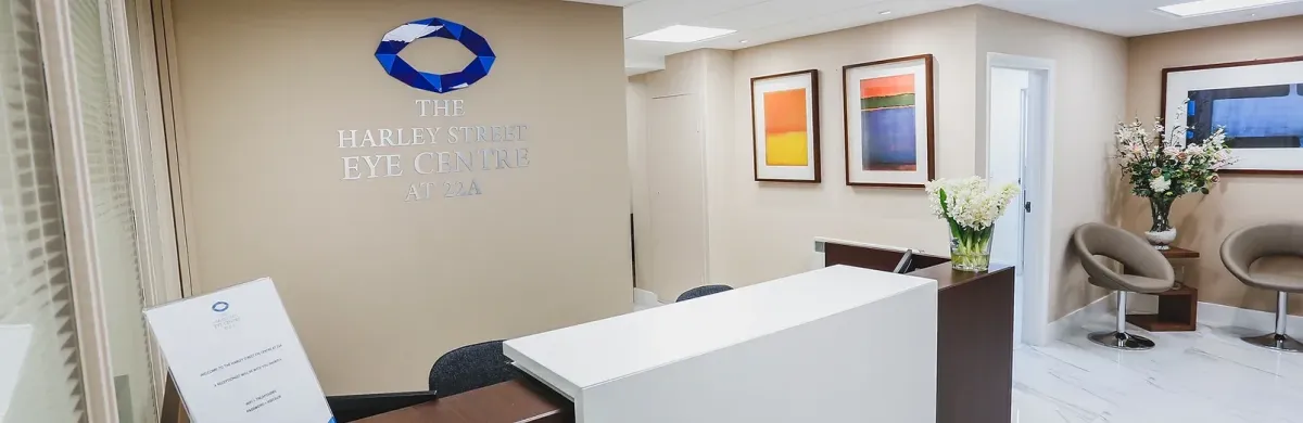
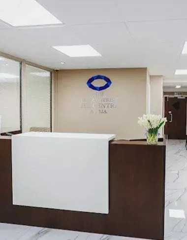
About Harley Street Eye Centre
Located in the heart of Harley Street in London, The Harley Street Eye Centre continues the decades-old association of eye care with 22a Harley Street. This brand new state of the art clinic offers full and comprehensive ophthalmic treatment and imaging. Medical Lead Mr. Marwan Ghabra and his team of leading ophthalmic surgeons and dedicated professionals are committed to providing world-class eye care.
The Harley Street Eye Centre provides the right eye treatment at the right time. Your eyes are precious, and you should take great care of them.
Centre for Complicated Cases
The Harley Street Eye Centre, in addition to providing high-quality eye care locally
in the UK, is quickly becoming well known internationally as a go-to centre for complicated cases needing out of the box thinking with a strong base in currently accepted ophthalmic practice.
When it comes to a routine eye test, a problem with squinting, double vision, loss of vision, a sore eye or unbearable eye pain or complex serious trauma to the eye, our team of experts are best suited to advise, examine and treat your eyes to bring you, improved eye health and peace of mind.
Located in The Heart of Harley Street London, UK
Here at The Harley Street Eye Centre, we have the knowledge and expertise to treat your eyes.
Questions you might be asking asking: where is there an eye specialist near me? Which eye clinic offers the best vision care in London? We're right here.
Located in the heart of Harley Street, near Marylebone in London we have undoubtedly the best solutions backed up by our team of medical experts. A consultation with one of our eye doctors will be your first step towards achieving the best possible outcome for your eyes.
We are not just a clinic, but a technologically advanced eye care facility filled with experts who collectively have decades of experience. A leading Eye Care provider in Harley Street, London, our experienced team of ophthalmologists, opticians and experts will find the best possible solutions all in one place.
The Harley Street Eye Centre continues the decades-old association of eye care with 22a Harley Street. Our brand new state of the art clinic offers full and comprehensive ophthalmic treatment and imaging using the latest technology.
Medical Lead Mr. Marwan Ghabra and his team of leading ophthalmic surgeons and dedicated professionals are committed to providing world-class eye care.
What We Treat
The Harley Street Eye Centre offers a comprehensive range of eye care services, catering to various eye health needs. From routine eye exams to advanced procedures such as refractive surgery (e.g., LASIK), cataract surgery, corneal transplants, and glaucoma treatment.
Corneal Treatment and Surgery
Keratoconus
Cataract Surgery
Lasik Laser Eye Surgery
Pediatric Eye Treatments and Surgery
TransPRK Laser Eye Surgery
Cosmetic Eye Surgery
Dry Eye Treatments
Retinal Surgery and Treatments
Glaucoma Eye Surgery and Treatments
From Consultation to Treatment
What To Expect At Your First Visit With Us
We're excited to see you at your appointment! We want to make sure you feel comfortable and well-informed, so don't hesitate to ask us any questions about our treatments, our office, or even our payment options. Our goal is to help you get back to living your life free of visual impairment, so let's work together to make that happen!
Discuss your eye health goals
Full eye examination
Vision check and treatment eligibility
Diagnostic scans if required
Start a custom treatment plan
"Highly recommend The Harley Street Eye Centre. The team are professional, experienced and educated."
- Subé W
The reviews showcased are authentic feedback from patients who have received treatment at The Harley Street Eye Centre. While individual outcomes may differ, these reviews do not claim to reflect the results of all patients.
Have Questions? Request a Call Back and Speak with a Doctor
One of our Doctors will call you for FREE and answer any questions or concerns you may have about your eye health
Fill in the form to request a call from one of our Doctors
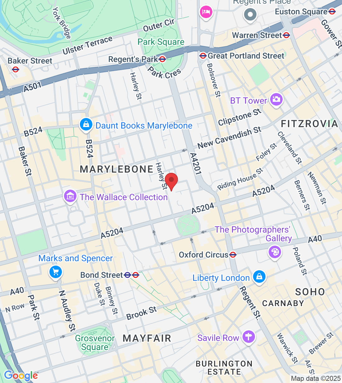
WHERE TO FIND THE HARLEY STREET EYE CENTRE
22a Harley Street, London W1G 9BP
Mon - Fri: 10am - 7pm
On street parking paid through ring go app, no free parking available
Bond Street and Oxford Circus Tube Station

© Copyright 2023. The Harley Street Eye Centre. All rights reserved.